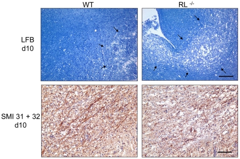
Figure 5. Increased demyelination and axonal damage in brain stem.
Brain stems of infected wt and RL−/− mice at day 10 p.i. stained with Luxol Fast Blue (top panels). Scale bar = 100 µm. Immunohistochemical staining for axonal integrity in brain stem at day 10 p.i. (bottom panels) Scale bar = 100 µm. Note more abundant loss of myelin and axonal degeneration in infected RL−/− relative to wt mice.

