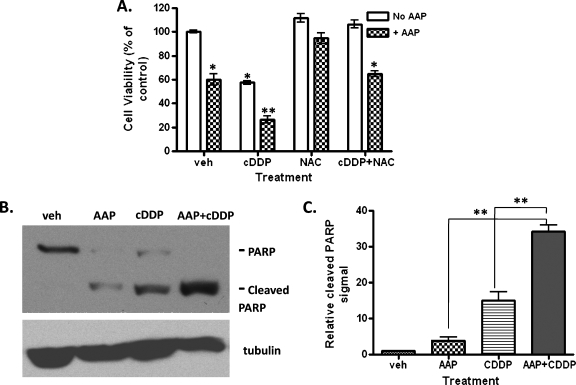
Figure 3.
Acetaminophen synergistically enhanced CDDP-induced cytotoxicity of HB cells in vitro. (A) HepT1 cells seeded in a 96-well plate were treated with 5 µg/ml CDDP, 10 mMAAP, and/or 1 mg/ml NAC and allowed to incubate overnight. Ten microliters of WST reagent was then added to each well, and the plate was incubated in normal culturing conditions at 37°C for 2 hours. The plates were read using a UV spectrometer at 450 nm. Results shown are normalized relative to the vehicle (n = 3, mean ± SEM). Statistical significance between treatment and control group was indicated by asterisks: *P < .05 or **P < .01. (B) Western blot analysis for levels of PARP and cleaved PARP was performed to measure apoptosis in the treated cells. Tubulin protein detection was used to confirm and normalize equal protein loading. (C) Quantification of the immunoblot signal of cleaved PARP in panel B was performed using UN-SCAN-IT Gel software (Silk Scientific) from three independent experiments. Statistical significance between two treatment groups was indicated: **P < .01.

