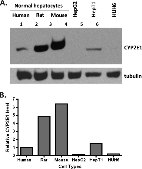
Figure 6.
Immunoblot analysis of CYP2E1 protein level difference in normal and malignantly transformed hepatocytes. Normal hepatocytes, hepatocarcinoma HepG2, and HB cells (HepT1 and HUH6) were obtained and prepared for immunoblot analysis as described in Materials and Methods section. (A) CYP2E1 protein level was measured using rabbit anti-CYP2E1 polyclonal antibody from Stressgen that cross-reacts with human, rat, and mouse. Antitubulin monoclonal antibody from Sigma that cross-reacts with human, rat, and mouse was used to confirm and normalize equal protein loading. (B) Quantification of the immunoblot signal of CYP2E1 presented in panel A was measured by normalization with tubulin protein level.

