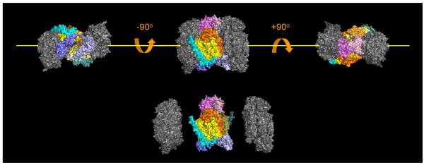
Figure 5.
Location of subunits within the three-dimensional structure of CcO that are either susceptible, or resistant to urea, or GdmCl-induced dissociation. The five subunits that dissociate (colored in this figure) are located near the dimer interface, and are in close contact with each other. This group of subunits is also the group that is sensitive to dissociation by elevated hydrostatic pressure [31]. Subunits that are resistant to dissociation, i.e., I, II, IV, Vb, VIc, VIIb, VIIc and VIII, are also clustered to form the catalytic core of CcO, (colored grey in this figure). Dimeric CcO can be considered to consist of three domains, two core domains that are urea and GdmCl resistant, and a third fragile, domain that bridges the other two (artificial separation of the CcO into these three domains is shown at the bottom of the figure). The third, fragile domain is roughly donut shaped with a hole in the center when viewed down the y-axis (yellow line). Dimeric CcO contains two copies of each subunit; therefore, the fragile domain is comprised of 10 subunits including two copies of subunits III (bright and lemon yellow), Vb (dark and light blue), VIa (bright and pale orange), VIb (bright and light pink), and VIIa (bright and dark cyan). All of the bright-colored subunits are associated with one CcO monomer, while all of the light, pale colored subunits are associated with the second monomer.

