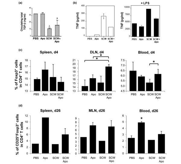
Figure 2.
Apoptotic-cell injection prevents streptococcal cell wall-induced arthritis by macrophageactivation prevention and regulatory T cells increase. (a) Rats from the different groups were punctured into the retro-orbital sinus at day 1 to quantify circulating total transforming growth factor beta (TGFβ) by ELISA in the serum (mean ± standard error of the mean (SEM); n = 3 rats per group, excepted PBS n = 2). Apo, apoptotic cells; SCW, streptococcal cell wall. Δ>P < 0.01 and *P < 0.05 compared with PBS-injected rats. (b) Macrophages issued from rats of the different groups were harvested from the peritoneum cavity 4 days after injection. TNF (mean ± SEM of the duplicate measurements) was tested by ELISA in the supernatant of the cultured macrophages (1 × 10⁶ cell per condition) from rats from each group (n = 3 to 4 rats) untreated (left panel) or after lipopolysaccharide (LPS) (50 ng/ml) overnight stimulation (right panel). Experiment repeated twice with similar results. (c) At day 4 and (d) at day 26 after SCW immunization, rats were sacrificed and the blood, spleen, inguinal lymph nodes (DLN) and mesenteric lymph nodes (MLN) were collected to analyze Foxp3+ regulatory T cells by flow cytometry. Results expressed as mean ± SEM; three animals/group; *P < 0.05. (c) Results for MLN and spleen expressed as mean ± SEM of the duplicate experiments, corresponding to two or three rats pooled together and repeated twice, not allowing statistical analysis. *P < 0.05 compared with PBS-treated or SCW-treated rats (four to six individual animals). (d) Experiment was repeated twice with similar results.

