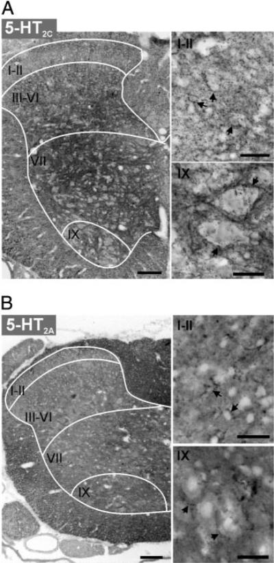
Fig. 3.
5HT2C and 5-HT2A receptor labeling in lumbar spinal cord of a P14 rat. For both 5-HT2C (A) and 5-HT2A receptors (B), left panel presents low power photomicrograph of one side of a spinal cord transverse section (scale bar = 100 μm) and right panels are confocal images displaying labeling at higher magnification (scale bar = 25 μm) of superficial dorsal horn (laminae I-II) and ventral horn lamina IX (top and bottom, respectively). Images are presented as grayscale negatives. Drawn white lines approximate white matter/ gray matter border and major divisions of Rexed's laminae. A: 5-HT2C receptor labeling is strongly expressed in the deep dorsal horn (lamina III-VI) intermediate lamina VII and motor nuclei (lamina IX). Arrows in right panels point to cell somas with perisomatic punctate labeling in dorsal horn (top) and lamina IX neurons in ventral horn (bottom). B: 5-HT2A receptor labeling is strongest in the white matter. Note that labeling in the gray matter is uniformly weak. Arrows point to punctate labeling that is not clearly juxtaposed to cell somata in the superficial dorsal horn (laminae I-II) and in the motor nucleus (lamina IX).

