
Figure 4.
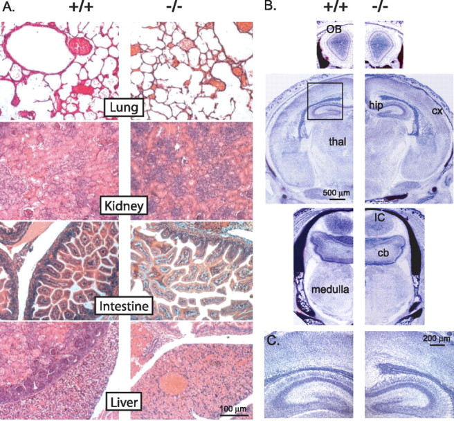
Normal GLS1 null mutant brain and peripheral development. A, Histopathological analysis (hematoxylin, eosin, safran staining) of newborn wild-type (+/+) and GLS1 null mutant (−/−) periphery showed no microanatomical differences. B, Photomicrograph showing Nissl-stained sections of forebrain, midbrain, and brainstem. No differences were observed in the brain size, gross structural organization, or ventricular size in GLS1 null (−/−) compared with wild-type mice (+/+). OB, Olfactory bulb; hip, hippocampus; cx, cerebral cortex; thal, thalamus; IC, inferior colliculi; cb, cerebellum. C, Enlargement of the rectangle in the second picture in C showing the normal cellular organization of hippocampal and cortical layers (cortical plate, layers V and VI) in GLS1 null mice.