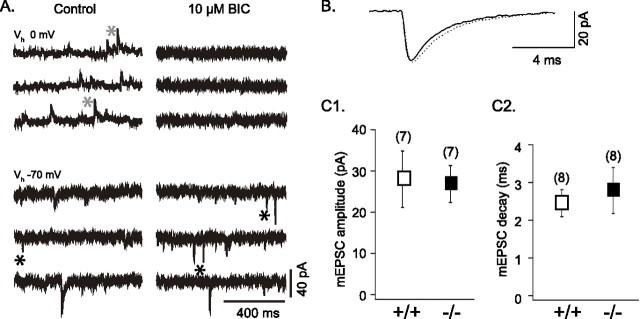
Figure 7.
Spontaneous EPSCs and IPSCs in GLS1+/+ and GLS1−/− cortical neurons in culture. A, In a GLS1−/− neuron (13 DIV), spontaneous synaptic currents were recorded under control conditions and after perfusion of 10 μm BIC at two holding potentials: at 0 mV, near the reversal potential of the EPSCs, and at −70 mV, near the resting potential. At 0 mV, the slow outward (upward) currents, recorded in control (gray asterisks) and blocked by 10 μm BIC, were GABAA-mediated IPSCs; they were also recorded at −70 mV as slow inward (downward) currents seen in control but not in BIC. The faster inward currents (black asterisks) that were recorded at −70 mV and persisted in BIC were spontaneous glutamatergic AMPA receptor-mediated EPSCs. B, Superimposed average of mEPSCs (n = 100), recorded in 1 μm TTX and 10 μm SR95531, from GLS1+/+ (black trace) and GLS1−/− (dotted trace) neurons showed no genotypic difference. C1, C2, The amplitude (C1) and the decay (C2) of the mEPSCs (measured in TTX and SR95531) showed no genotypic difference. Error bars represent SD.

