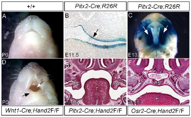
Figure 4.
Epithelial specific inactivation of Hand2 leads to cleft palate formation. (A) A newborn wild type mouse shows a normal mandible. (B) A section through oral cavity of an E11.5 Pitx2-Cre;R26R embryo shows specific LacZ staining in the palatal epithelium (arrow) and dental epithelium (de). (C) Whole mount LacZ staining of an E13.5 Pitx2-Cre;R26R embryo demonstrates Cre activity in the entire palatal shelf (arrows). (D) A newborn Wnt1-Cre;Hand2LoxP/LoxP mouse (labeled as Wnt1-Cre;Hand2F/F) exhibits a hypoplastic mandible (black arrow) and an intact palate (white arrow). (E) A section through oral cavity of an E16.5 Pitx2-Cre;Hand2LoxP/LoxP embryo (labeled as Pitx2-Cre;Hand2F/F) shows cleft palate defect. Note the palatal shelves remain in a vertical position. (F) A section through oral cavity of an E16.5 Osr2-Cre;Hand2LoxP/LoxP embryo (labeled as Osr2-Cre;Hand2F/F) shows a normal formed palate. T, tongue; de, dental epithelium; PS, palatal shelf.

