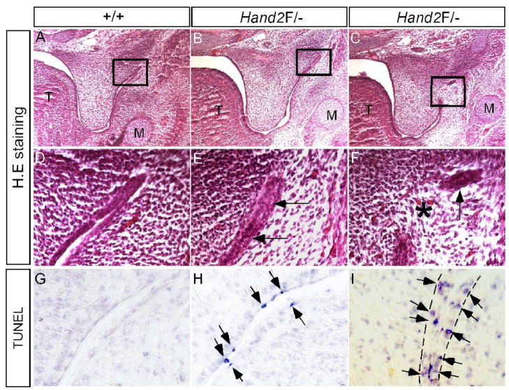
Figure 7.
Pathological adhesion/fusion of the palatal shelf and mandible in Hand2 hypomorphic mice. (A, D, G) E13.5 wild type controls show normal histological structure (A, D) and absence of apoptotic cells in the lateral junction of the posterior palate and mandible. (B, E) An E13.5 Hand2LoxP/− embryo shows adherence of epithelia (arrows in E) of the lateral junction of the posterior palate and mandible. (C, F) An E13.5 Hand2LoxP/− embryo shows fusion of the palatal shelf and mandible at the lateral junction. Star denotes a confluence of mesenchyme, and arrow points to a remanent epithelium. (H) An E13.5 Hand2LoxP/− embryo shows apoptotic periderm cells (arrows) in the lateral junction right before epithelial contact/adherence. (I) An E13.5 Hand2LoxP/− embryo shows apoptotic basal layer epithelial cells (arrows) in the lateral junction fusion of the palatal shelf and mandible. The dash lines demarcate the epithelial boundary.

