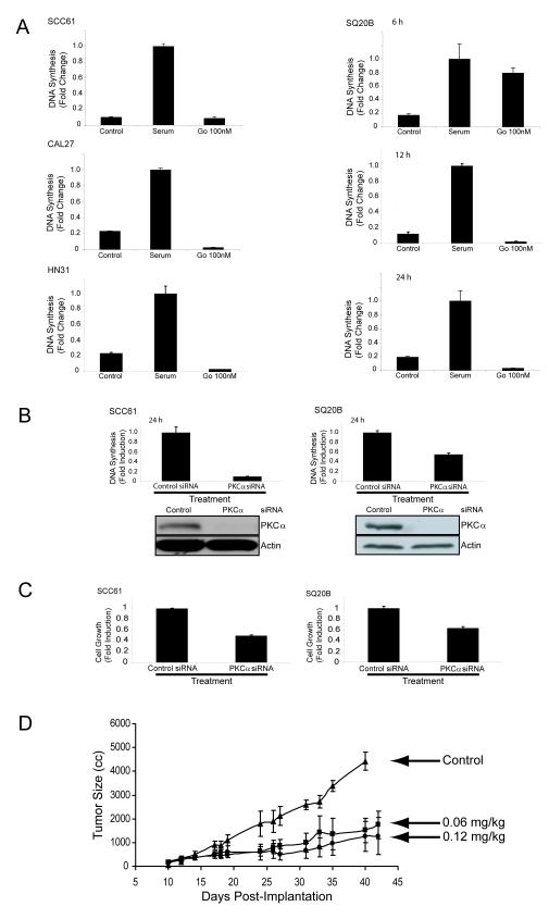
Figure 1. PKC alpha inhibition reduces DNA synthesis, cell viability, and tumor growth in vivo.
A) SCC61, SQ20B, CAL27, and HN31 cell lines were incubated in serum-free media (control), serum alone (serum), or serum with 100 nM of Gö6976 (Gö 100nM) for up to 24 hours and DNA synthesis was quantitiated for BrdU incorporation as described in Experimental Procedures. B) SCC61 (left) and SQ20B (right) cells were transfected with control or PKC alpha siRNA. DNA synthesis was quantitiated for BrdU incorporation as described in Experimental Procedures. All BrdU incorporation results are the mean +/- S.E. of three independent experiments. Immunoblotting for PKCα and actin was performed in conjunction with BrdU incorporation after control and PKCα siRNA transfection as described in Experimental Procedures. C) SCC61 (left) and SQ20B (right) cells were transfected with control or PKCα siRNA and cell survival was quantitated by MTT assay as described in Experimental Procedures. All MTT results are the mean +/- S.E. of three independent experiments. D) SQ20B tumor xenografts were established in female athymic nude mice. After reaching an average size of 150 mm3 the mice (n = 5) were treated with 0.1% DMSO in PBS (control) or Gö6976 (n = 10 per group) at 0.06 mg/kg/week or 0.12 mg/kg/week by intravenous tail vein injection. Values represent mean tumor size +/- S.E.

