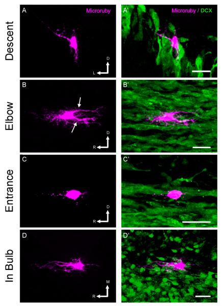
Figure 5. Reconstructions of astrocytes in the RMS.
Individual astrocytes in the RMS of DCX-GFP mice were filled by intracellular injection of rhodamine- and biotin- conjugated dextran (magenta) in fixed tissue slices, imaged, and 3D reconstructed. Left panels show volume reconstructions of the astrocyte, and right panels show the relationship of the filled cell to the DCX+ chains of neuroblasts (green). Note the polarity of the astrocytes, with long processes oriented parallel to the direction of migration (arrows). In C, the endfeet of the astrocyte can be seen along a migrating chain. Scale bar: 25 μm. R: rostral; D: dorsal; L: lateral; M: medial. The orientation of tissue sectioning was coronal in A, sagittal in B and C and horizontal in D.

