
Figure 5. The combination of fractionated radiotherapy with anti-CTLA-4 antibody enhances TIL in secondary TSA tumors and tumor-specific T cells producing IFNγ.
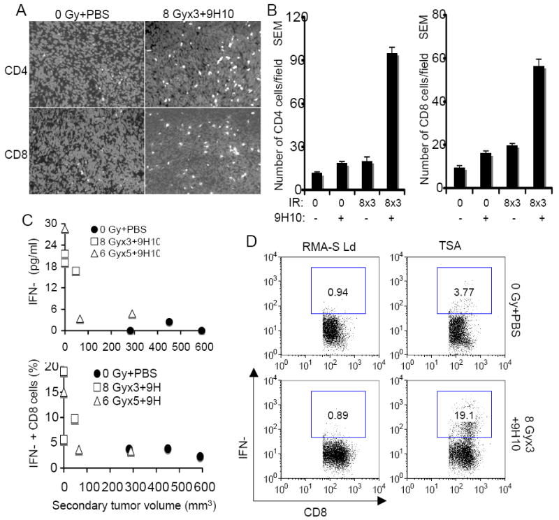
(A, B) Secondary tumors were excised at day 35 and analyzed by fluorescence microscopy for the presence of CD4+ and CD8+ T cells. (A) Representative fields showing CD4+ (top panels) and CD8+ (bottom panels) T cells (white) infiltrating secondary TSA tumors in mice treated as indicated. Nuclei were stained with DAPI (light gray). (B) Mean number ± SE of CD4+ and CD8+ TILs in three mice per group. Both CD4+ and CD8+ TIL were significantly increased in mice treated with the combination of 8 Gy × 3 + 9H10 (p<0.05 compared to all other groups), whereas radiation and 9H10 as single modalities did not have a significant effect. (C, D) Analysis of tumor-specific IFNγ production by spleen cells harvested at day 35 from mice in the various treatment groups. (C) IFNγ concentration in supernatants of total spleen cells isolated from mice treated with 0 Gy + PBS (closed circles), 8 Gy × 3 + 9H10 (open squares), or 6 Gy × 5 + 9H10 (open triangles) and cultured o.n. with irradiated TSA cells were plotted against the volume of the secondary tumor (Top panel). The percentage of CD8+ T cells expressing IFNγ when exposed to TSA cells as determined by intracellular staining (D) following in vitro restimulation with the TSA-derived immunodominant CD8 epitope AH1 was plotted against the volume of the secondary tumor (Bottom panel). Symbols are as above. Each symbol represents one animal. (D) Representative histograms showing the percentage of CD8+ T cells positive for IFNγ by intracellular staining and flow cytometry in response to TSA cells or the irrelevant target RMA-S-Ld. Samples were gated on CD8+ T cells.