
Figure 6. The abscopal effect is induced in MCA38 tumor-bearing mice by fractionated radiation in combination with anti-CTL-4 antibody.
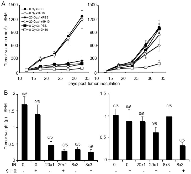
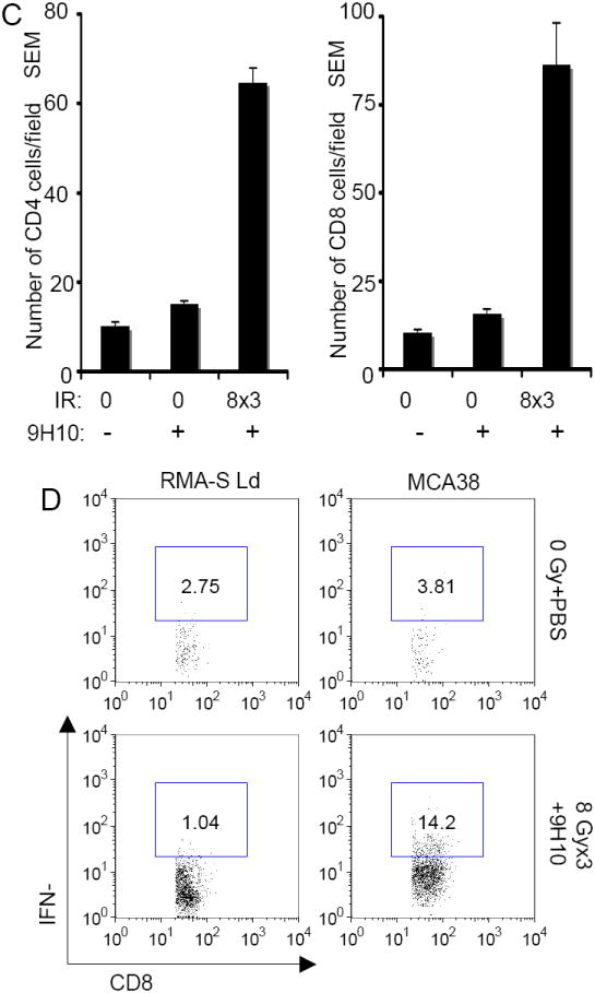
C57BL/6 mice were injected with syngeneic MCA38 colon carcinoma cells (5 × 10⁵) s.c. into the right and left flank as outlined in Figure 1. (A) Tumor growth delay of primary irradiated tumors (left panel) and secondary non-irradiated tumor (right panel) in mice treated with PBS (closed circles), 9H10 (open circles), 20 Gy × 1 + PBS (closed diamonds), 20 Gy × 1 + 9H10 (open diamonds), 8 Gy × 3 + PBS (closed squares), 8 Gy × 3 + 9H10 (open squares). 9H10 was given on days 14, 17, and 20. Data are the mean ± SE of 5 mice/group. (B) Tumor weight of primary (left panel) and secondary (right panel) tumors at day 35. Data are the mean ± SE. The number of mice with complete tumor regression over the total number of mice per group is indicated. (C) Secondary tumors were excised at day 35 and analyzed by fluorescence microscopy for the presence of CD4+ and CD8+ T cells. Data are the mean number ± SE of CD4+ and CD8+ TILs in three mice per group. Both CD4+ and CD8+ TIL were significantly increased in mice treated with the combination of 8 Gy × 3 + 9H10 (p<0.05 compared to all other groups). (D) Analysis of tumor-specific IFNγ production by spleen cells harvested at day 35 from treated and untreated mice and re-stimulated in vitro with irradiated MCA38 cells. Histograms show the percentage of CD8+ T cells positive for IFNγ by intracellular staining and flow cytometry in response to MCA38 cells or the irrelevant target RMA-S-Ld. Samples were gated on CD8+ T cells. Spleen cells from 3 mice in each treatment group were pooled.