
Figure 12.
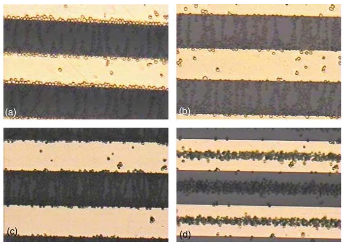
(online color at: www.biophotonics-journal.org) Bands of yeast cell aggregations under 8 Vp-p at sinusoidal frequencies of a) 10 kHz and b) 5 MHz for viable yeast cells and c) 10 kHz and d) 5 MHz for nonviable yeast cells. The bright and dark regions are electrodes and glass substrate respectively, and the cells are immersed in a 5 μS/cm buffer fluid. Reprinted from [94] with permission from Elsevier.