
Figure 5.
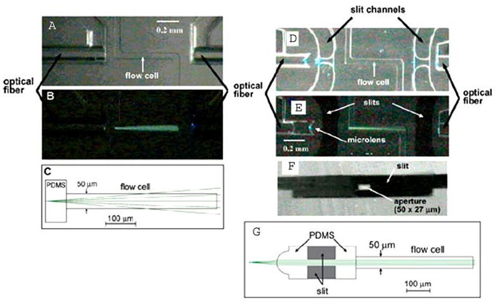
(online color at: www.biophotonics-journal.org) Left: Ro et als Flow cell (a) illuminated only by a fiber optic (b), exhibiting divergence predicted by ray-tracing simulations (c). Right: A similar flow cell (d) with a lensed air space (microlens) at the end of the fiber. The microlens acts to collimate the light from the fiber (e) with the help of stray light blocking by the aperture (f), as expected from ray tracing simulations (g). The resulting smaller, more uniform interrogation beam would be desirable for uniform, localized excitation in flow cytometry chips. Reprinted with permission from [56].