Fig. 8.
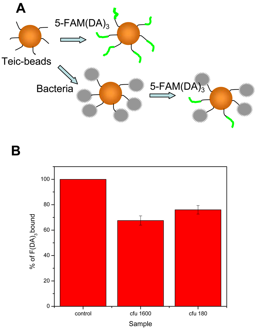
Gram-positive bacteria binding to teic-bearing magnetic microspheres. (a). Schematic diagram showing the basis of bound bacteria determination. Teic-microspheres will be incubated with 1 in control. Teic- microspheres will be incubated with bacteria first and then with 1 for test samples. (b). Degree of bacteria binding is determined by the percentage of fluorescence decrease compared to the control.
Gram positive bacteria binding to teicoplanin bearing magnetic beads. A. Schematic diagram showing the basis of bound bacteria determination. Teic-beads will be incubated with 5-FAM(DA)3 in control. Teic-beads will be incubated with bacteria first and then with 5-FAM(DA)3 for test samples. B. Degree of bacteria binding is determined by the percentage of fluorescence decrease with comparison to control.

