Fig. 6.
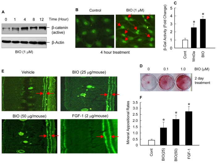
BIO activates β-catenin signaling stimulates osteoblast differentiation and bone formation. Primary calvarial osteoblasts were isolated 3-day-old WT neonatal mice and cultured with or without BIO (1 μM) for different periods of time as indicated. The β-catenin protein levels and nuclear translocation were examined by western blot and immunostaining. BIO increased protein levels of active form of β-catenin and reached its maximal effect at 4 hours (A). BIO also induced β-catenin nuclear translocation (B). Primary calvarial osteoblasts, isolated from TOPGal transgenic mice, were treated with BIO (1 μM) for 24 hours and β-Gal activity was measured using cell lysates isolated from these cells. Wnt3a (100 ng/ml) was used as a positive control in this experiment. BIO significantly increased the β-Gal activity (C). *P<0.05, unpaired Student's t-test, n=3. Primary calvarial osteoblasts were cultured with 0.1 and 1 μM of BIO for 2 days and changes in ALP activity were examined by ALP staining. BIO (at both concentrations) significantly increased ALP activity in these cells (D). To further determine whether BIO induces new bone formation in vivo, 25 and 50 μg/mouse of BIO was injected into 1-month-old WT mice subcutaneously over the surface of calvariae for 5 days. Calcein labeling was performed at day 5 and 15. Mice were sacrificed 2 days after the second calcein labeling and periosteal new bone was evaluated and mineral appositional rates (MAR) were measured. FGF-1 (2 μg/mouse, 5 day injection) was used as a positive control. BIO significantly increased new bone formation and MAR in this assay (E,F). *P<0.05, one-way ANOVA followed by Dunnett's test (BIO versus control) and unpaired Student's t-test (FGF-1 versus control), n=5. All values are means ± s.e.

