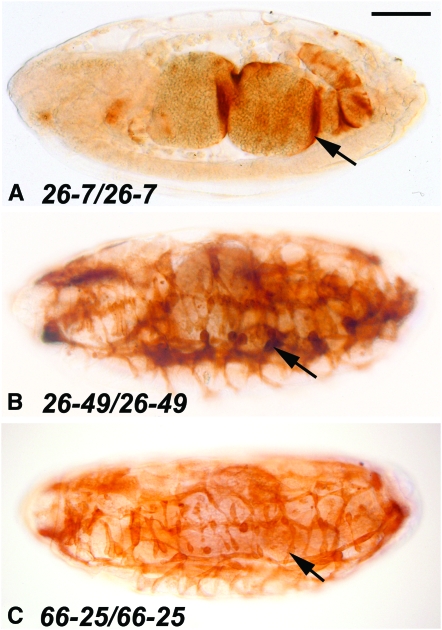
Figure 6.—
Skeletal muscle development in three new Mef2 mutant alleles. Stage 16 embryos raised at 29° were stained with anti-tropomyosin to visualize skeletal muscle patterning and differentiation. All embryos are sagittal views with anterior to the left. Embryos of the same genotypes raised at 18° showed similar phenotype to those shown here. (A) The 26-7 homozygous mutant showed a Mef2 null phenotype, since there was no skeletal muscle development detected, and only a small amount of stain was observed in the visceral muscle surrounding the gut (arrow). (B and C) The 26-49 and 66-25 homozygous mutants showed some level of skeletal muscle development (arrows); however, the phenotypes overall were indicative of these each being strongly hypomorphic alleles. Bar, 100 μm.

