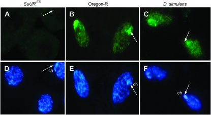
Figure 2.—
Immunodetection of SUUR in follicular cells with E45 antibodies. (A) Negative control: no staining is observed in follicle cells of SuURES. (B) Positive control: strong staining is observed in the nucleus and especially the chromocenter (ch, indicated by arrows) in follicle cells of D. melanogaster (Oregon-R). (C) In D. simulans, the antibodies produce staining similar to that observed in Oregon-R. (D–F) Hoechst staining.

