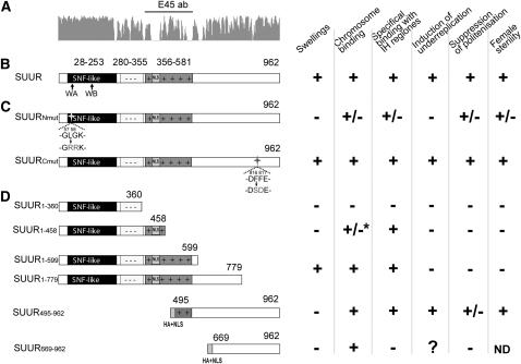
Figure 3.—
Organization and features of different SUUR isoforms. (A) Conservation plot, based on the SUUR sequences from 11 Drosophila species, and the protein region used to generate E45 antibodies (E45 ab). (B) Domain organization of SUUR protein. The SNF-like domain is solid, the negatively charged region is marked with - -, and the positively charged amino acid region is darkly shaded with + +. NLS, nuclear localization signal. Arrows show the position of regions with similarity to Walker A (WA) and Walker B (WB) motifs of SNF2/SWI2 proteins. (C) Point mutations introduced within the N- and C-terminal part of SUUR are indicated by arrows. (D) Truncated fragments of SUUR are described by Kolesnikova et al. (2005) except for SUUR669–962. HA+NLS, hemagglutinin tag and nuclear localization signal sequence. An asterisk indicates that granules in intercalary and pericentric heterochromatin were observed.

