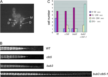
Figure 2.—
Myo1 ring contraction is defective in the bub3 clb5-1 mutant. (A) Cells were incubated in YPD liquid media at 23° and put on SC-complete media containing agarose at low density. A movie was taken at 37° as described in materials and methods. The image is from the picture after 8.75 hr at 37°. M indicates mother cells, and D indicates daughter cells. (B) Myo1-GFP images at the bud neck were taken every 30 sec with time-lapse fluorescent microscopy. The images were trimmed to focus on the bud neck area, and at least 40 images were put next to each other to follow the myosin ring contraction. (C) The time length of Myo1-GFP expression on the bud neck was analyzed in the time-lapse images. Ten cells for each genotype were monitored to monitor how many seconds Myo1-GFP stayed on the bud neck.

