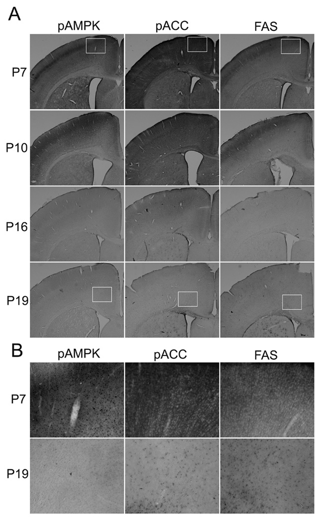
Fig. 2.

Developmental changes in immunostaining patterns of brain sections labeled with antibodies against pAMPKα (Thr172), pACC (Ser79), and FAS. In Fig.2A, brain sections from P7, P10, P16, and P19 mice were immunostained with anti-pAMPKα (Thr172) antibody, anti-pACC (Ser79) antibody or anti FAS antibody using the ABC method as described in Experimental Procedure. The size of a rectangle inserted into panels of P7 and P19 is 0.59 mm × 0.47 mm. Fig. 2B shows magnified images of the area indicated as rectangles in Fig. 2A.
