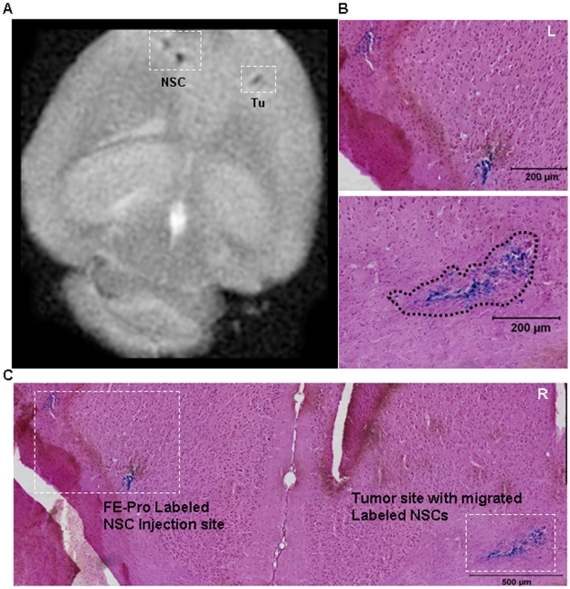
Figure 5. Sensitivity of MRI monitoring of FE-Pro-labeled NSCs targeting human glioma.
(A) T2-weighted MR image of mouse brain in Fomblin, showing two distinct signal voids generated by FE-Pro-labeled NSCs that were injected ∼200 µm apart from each other on the left hemisphere and a hypointense signal generated by FE-Pro-labeled NSCs that migrated to the contralateral tumor site (white dotted boxes). Approximately 600 FE-Pro-labeled NSCs constituted a detectable signal void. (B and C) Prussian blue stained section from the region shown in (A). Higher magnification images (B, tumor area denoted by black dotted line) of the regions outlined in (C), showing PB-positive labeled NSCs corresponding to the hypointense signal sites in (A). MRI conditions: 7.0 Tesla, Rapid Acquisition Relaxation Enhancement sequence, 78 µm/pixel, 300 µm/slice, TR/TE = 1500/23.1 ms. Scale bars = 200 µm (B), 500 µm (C).

