Figure 1.
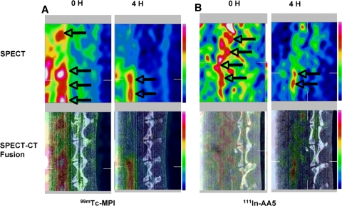
Noninvasive radionuclide imaging of Tc-MPI and In-AA5 in atherosclerosis. Micro-SPECT (top), and micro-SPECT-CT fusion images (bottom) of atherosclerotic rabbit on uninterrupted HC diet using Tc-MPI (A) and In-AA5 (B). The left sets in A and B display images immediately (0 hour) after radiotracer administration, which reveal blood pool activity in the aorta (arrows) in front of vertebral column. The right sets in A and B show target localization 4 h after radiotracer administration. The radiotracer uptake in the atherosclerotic lesions of the abdominal aorta is observed (arrows)

