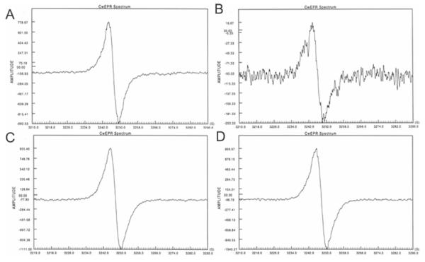
Figure 4.
EPR analyses of mycelial S. schenckii melanin particles generated on minimal medium (A), minimal medium with 8.0 mg/L tricyclazole (B), minimal medium with L-DOPA (C) and minimal medium with tricyclazole C and L-DOPA (D). Note that amplitude of signals is lower on (B) and higher on (C) and (D). The spectrum on (B) was recorded at maximum gain in an effort to identify a signal and hence has more background noises. Acid-resistant S. schenckii particles derived from yeast forms had similar spectra to mycelial particles generated from the same culture medium.

