
Figure 1. Interaction of ATL1 with Rtn4a in neuronal cells.
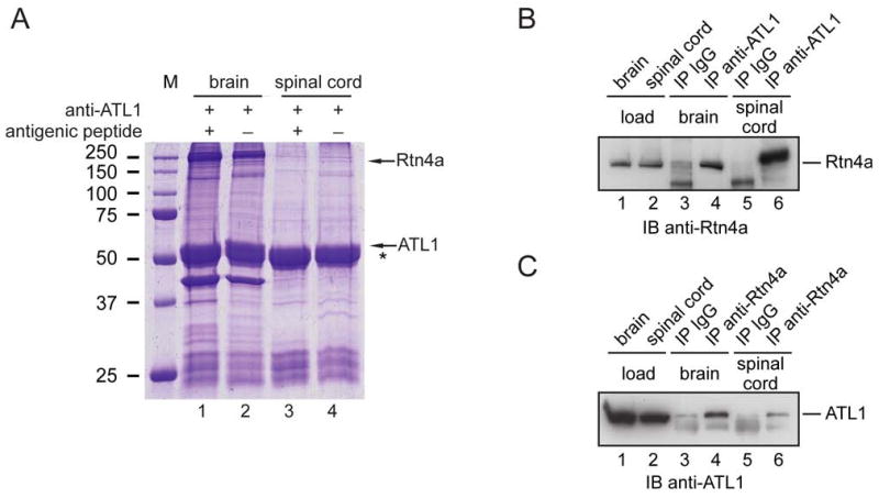
(A) Detergent extracts from mouse brain or spinal cord were incubated with peptide-specific antibodies to ATL1. Where indicated, antibodies were preincubated with the antigenic peptide. Immunoprecipitated proteins were analyzed by SDS-PAGE and Coomassie staining. Rtn4a was identified by mass spectrometry (5 distinct peptides covering 74 of the 1163 amino acids). An asterisk (*) indicates the position of the IgG heavy chain. M, molecular mass standards (in kDa).
(B) Proteins immunoprecipitated (IP) with ATL1 antibodies or control IgG were analyzed by immunoblotting (IB) with Rtn4a antibodies. Lanes 1 and 2 (loads) show 10% of the starting material used for immunoprecipitation.
(C) Proteins immunoprecipitated with Rtn4a antibodies or control IgG were immunoblotted with ATL1 antibodies. Lanes 1 and 2 (loads) contain 10% of the starting material used for the immunoprecipitations.