
Figure 7. Sey1p and Rtn1p/Yop1p cooperate in maintaining ER morphology in S. cerevisiae.
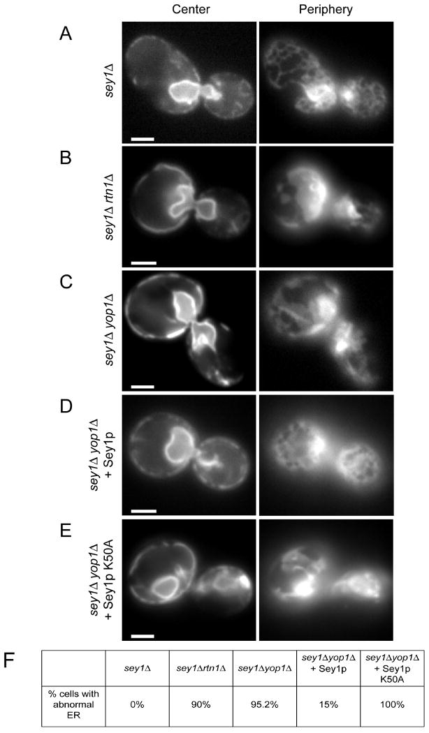
(A) A GFP-fusion of the ER protein Sec63p was expressed in cells lacking Sey1p (sey1Δ). The localization of the protein was determined by fluorescence microscopy, focusing the microscope either at the center or the periphery of the cell. Bar, 1 μm.
(B) As in (A), but with cells lacking both Sey1p and Rtn1p (sey1Δrtn1Δ).
(C) As in (A), but with cells lacking both Sey1p and Yop1p (sey1Δyop1Δ).
(D) As in (C), but with cells expressing wild-type Sey1p from a CEN plasmid.
(E) As in (C), but with cells expressing a GTP-binding mutant of Sey1p (Sey1p K50A) from a CEN plasmid.
(F) The percentage of cells with abnormal ER was determined from 20-40 cells per mutant. The small percentage of cells with abnormal ER in (D) likely reflects loss of the plasmid.