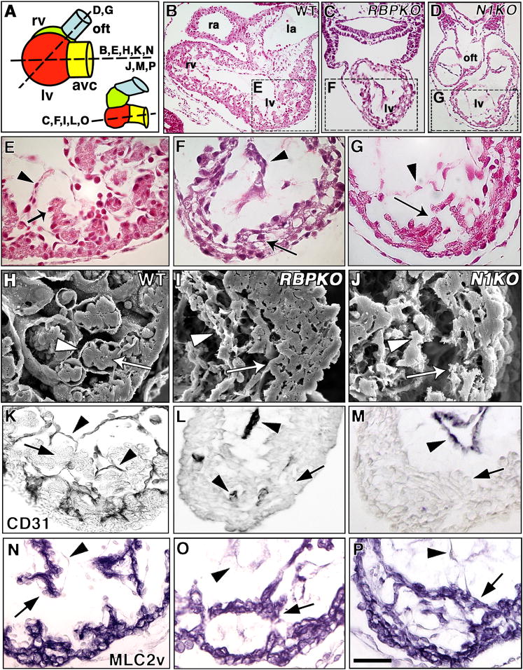
Figure 2. Defective trabeculation in E9.5 Notch1 and RBPJk mutants.
(A) Schematic representation of E9.5 wt or Notch1 (left) and RBPJk mutant hearts (right), depicting a lateral view of cardiac chambers. Section levels shown in (B–P) are indicated. In RBPJk mutants, the ventricles are aligned along the A–P axis. avc, AV canal; oft, outflow tract. (B–G) H+E-stained transverse sections and (H–J) SEM photomicrographs. (B, C, D) General views of representative wt (B), RBPJk (C) and Notch1 (D) mutants at the level of ventricles. Details of wt left ventricle (E, H), note developing trabeculae with myocardium (E, H, arrows) and endocardium (arrowheads); left ventricles of RBPJk (F, I) and Notch1 (G, J) mutants. RBPJk and Notch1 embryos show collapsed endocardium (arrowheads), disorganized myocardium (arrows) and poorly developed trabeculae. (K–M) Endocardial CD31/PECAM staining (arrowheads) in the left ventricle of wt (K), RBPJk (L) and Notch1 (M) embryos. In wt trabeculae the myocardium (arrow) is surrounded by endocardium (arrowhead). Arrowheads in (L, M) show endocardium and arrows indicate myocardium. (N-P) MLC2v staining in wt (N), RBPJk (O) and Notch1 (P) embryos show MLC2v expression throughout myocardium including trabeculae (arrow). Arrowheads indicate endocardium. la, left atrium; oft, outflow tract; ra, right atrium. Scale bar, 100 μm in B–D; 25 μm in D–P.

