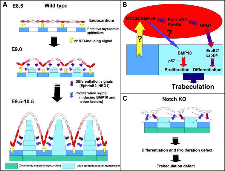
Figure 7. A model for Notch activity in ventricular development.
(A) E8.5 wt embryo. A myocardial cue (yellow) leads to N1ICD expression (red) in specific endocardial regions. At E9.0, N1ICD/RBPJK activate endocardial EphrinB2 leading to NRG1 expression. NRG1 activates the ErbB2/B4 receptors in myocardium to induce trabecular muscle differentiation. As the trabecular ridge develops, the endocardium separates from the myocardial N1ICD-inducing cue, and N1ICD is down-regulated (pink) at the tip of the trabeculae. At E9.5-10.5, N1ICD is higher in the endocardium at the base of the trabeculae, and the ventricular myocardium has differentiated into compact zone (green) and trabecular myocardium (blue) regions. The different intensity of N1ICD expression (pink or red) represents the predominant N1ICD activation at the base of trabeculae in response to a spatially restricted myocardial cue. (B) Molecular pathways downstream of Notch during trabeculation. (C) In Notch mutants, proliferation and differentiation signals are disrupted and trabeculation is impaired.

