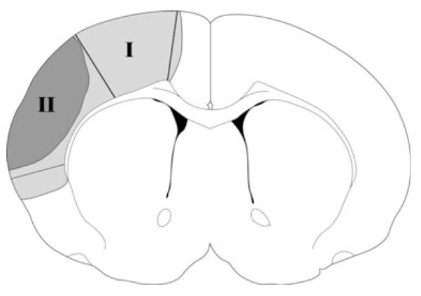
Fig. 1.

Tissue corresponding to the ischemic penumbra and core. The gray region (I), including the black region (II) represents ischemic injury in controls with ischemia alone; the black region (II) represents infarction in ischemia plus postconditioning. The region spared by postconditioning is defined as the penumbra (region I) and the black area is defined as the ischemic core (region II). These regions were dissected for western blotting. The corresponding non-ischemic cortex from sham animal without ischemia was dissected for comparison.
