
Figure 1.
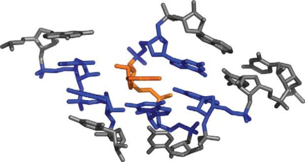
A section of the large ribosomal subunit (PDB ID 1JJ2) is shown, with a single nucleotide colored orange. This nucleotide forms a separate nucleotide doublet with each of the five residues colored blue. In the set of structures we use here, the average nucleotide is part of five doublets. There are some nucleotides that are part of just one doublet and a very small number of nucleotides that are part of 12 different doublets.