
Figure 2.
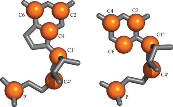
At left a guanine and at right a cytosine nucleotide, oriented in similar fashion. Highlighted in orange are atoms involved in RMSD calculations. There are six atoms in all, three in the backbone (P, C4′ and C1′) and three in the base (C2, C4 and C6). The different orientation of the six-membered ring between purines and pyrimidines serves to easily differentiate between the two types of nucleotide, and the distribution of atoms provides good sampling of both the base and sugar-phosphate backbone.