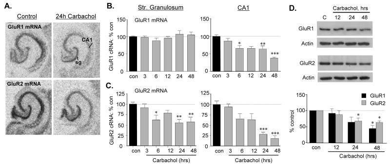
Figure 3.
GluR mRNA and protein levels are reduced by carbachol treatment. A) In situ hybridization labeling of GluR1 and GluR2 mRNAs in control and carbachol-treated (50 μM, 24 h) hippocampal slices: carbachol decreased both mRNAs with effects being greatest in CA1 str. pyramidale (abbreviations: sg, str. granulosum; CA1, field CA1 str. pyramidale). B and C) Bar graphs show quantification of hybridization densities to GluR1 (B) and GluR2 (C) mRNAs in str. granulosum and CA1 str. pyramidale after 3 to 48 h treatments (mean ± SEM values presented as percent paired control; *p < 0.05, **p < 0.01, ***p< 0.001 vs. con, SNK; n ≥ 10–28 and 7–10 slices/group for GluR1 and GluR2 mRNAs, respectively). D) Representative western blots and quantification of band densities (normalized to same-sample actin-ir, presented as percent paired control values) show total GluR1 and GluR2 protein levels in cultured hippocampal slices harvested without treatment (C, con), or after 12 to 48 h 50 μM carbachol treatment (*p < 0.05 versus control, SNK; n ≥ 4/group). Actin-ir was unaffected by carbachol treatment (D).

