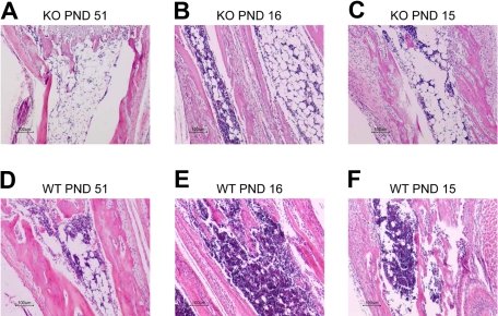
Figure 5.
Histologic analysis of bone marrow in Zfp36l2 KO and WT mice. Tissue sections from the radius/ulna of PND 51 KO (A) and WT (D) mice, PND 16 KO (B) and WT (E) mice, and PND 15 KO (C) and WT (F) littermate pairs of mice were stained with hematoxylin and eosin. Note the hypocellular bone marrow along with an increased number of adipocytes in the KO samples. Original magnifications ×10, obtained with a Nikon Eclipse E600 microscope with a Nikon DXM1200 digital camera. Images were imported into the Nikon Act-1 software.

