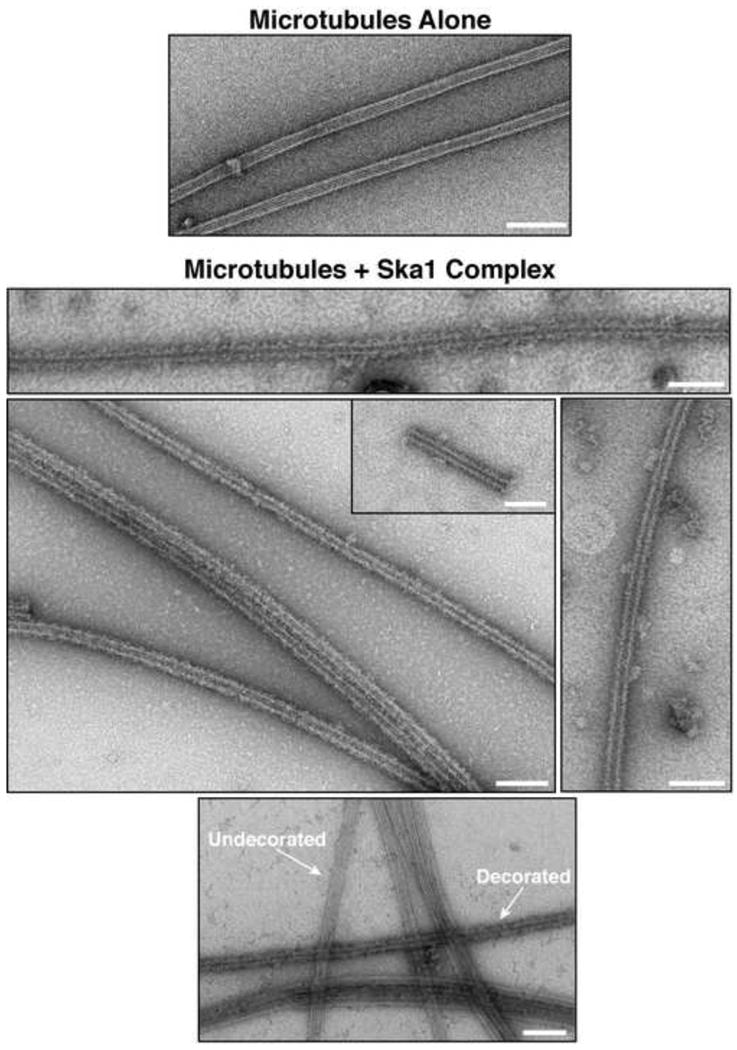
Figure 5. The Ska1 complex forms ring-like assemblies on microtubules.

Electron microscopy of negatively stained taxol stabilized microtubules either alone or with Ska1 complex bound. In the bottom image, microtubules with Ska1 complex bound and undecorated microtubules are visible in the same field (indicated by arrows). Scale bars, 100 nm.
