
FIGURE 2.
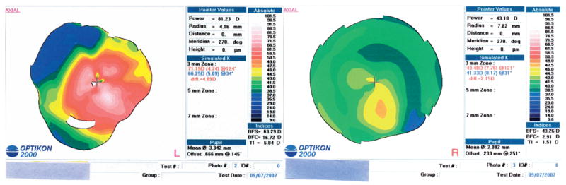
Case 2 with keratoconus (topographies after INTACS). Corneal topography of both eyes before corneal transplantation. Left eye (L), severe inferonasal steepening involving the visual axis. There is an irregular asymmetric bowtie centered on the visual axis; right eye (R), inferior steepening with asymmetric and skewed bowtie. There is a curved area of flattening in the nasal midcornea corresponding to the nasal INTACS.