
FIGURE 3.
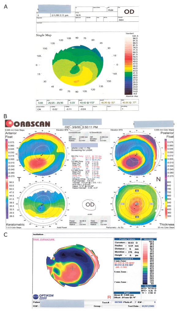
Case after LASIK keratectasia topographies. A, Corneal topography of the right eye before LASIK. A typical crab claw pattern is visible, suggesting the presence of pellucid marginal degeneration preoperatively. B, Corneal topography of the right eye after LASIK. Orbscan shows abnormal anterior and posterior floats, and the keratometric tracing shows again a pattern consistent with pellucid marginal degeneration. C, Corneal topography of the right eye after INTACS and before corneal transplantation. It shows an area of inferior steepening involving the optical center and the lack of the crab claw pattern previously observed.