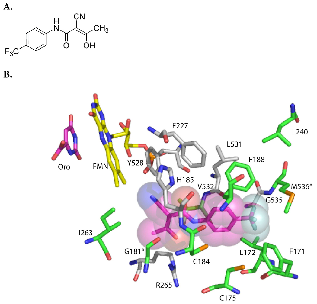
Figure 1. Species-variable inhibitor binding site of PfDHODH.
A. Structure of the DHODH inhibitor 2-cyano-3-hydroxy-N-[4-(trifluoromethyl)phenyl]-2-butenamide (A77 1726) 1. B. Amino acid residues within 4.2 Å of the co-crystallized inhibitor 1 are shown. The inhibitor (pink) is displayed showing the Van der Waals surface, the FMN cofactor (yellow) and orotate (pink) are displayed as sticks. Residues in grey are conserved between PfDHODH and hDHODH, while residues in green are variable. The two amino acid residues that differ between PfDHODH and PbDHODH are indicated by *. The figure was generated using PyMol from the file 1TV5.pdb.

