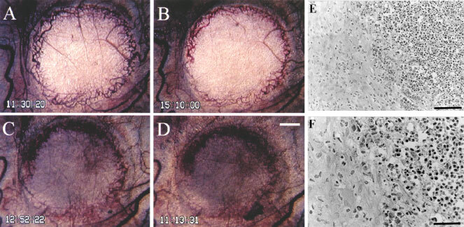
Figure 8.

Typical finding of growth inhibition caused by AC7700 in an SLC microtumour developing in a transparent chamber. (A) before administration of 10 mg kg−1 AC7700 administration; (B) 3.5 h after administration of AC7700; (C) 25 h later; (D) 48 h later; (E) histology 48 h later. Tumour blood flow completely stopped at 1 h after a single i.v. administration of AC7700. The whole region of the tumour, with a diameter of 2.5 mm, became necrotic. Tumours stopped growing completely during the 48-h observation period. Histological study (E and F, H&E stained) certified the tumour (shown on the right side) as necrotic. Original magnification: A–D, ×20; E, ×200; F, ×400. Bars: A–D, 500 μm; E, 100 μm; F, 50 μm.
