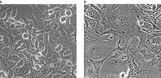
Figure 1.

Typical morphological differences between B16 F0 melanoma cells that were frozen in the absence (A) or in the presence of 100 nm of bleomycin (B), after 137 h of incubation. (Magnification 1100×). In the absence of bleomycin, the surviving cells display a normal morphology, regular cell size, and the usual nucleus aspect. Normal mitosis can be observed in the two rounded cells on the right upper part of panel A. In contrast, in panel B cells show abnormal morphology. The size of the nuclei and cytoplasm has increased. The limits of the nucleus are not always demarcated while the cytoplasm itself is clear. Among cells there are large differences in nuclei and cell size.
