
Figure 3.
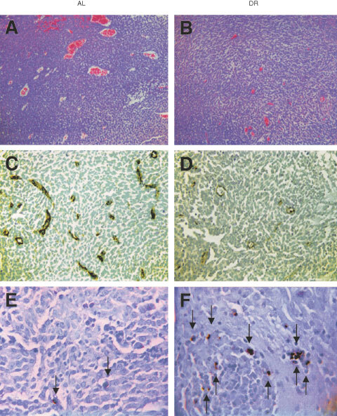
Influence of DR on microvessel density and apoptosis in the CT-2A brain tumour. DR was initiated 7 days before intracerebral tumour implantation and was continued for 11 days. H&E stained tumour sections in an AL mouse (A) and in a DR mouse (B) (100×). Factor VIII immunostaining from the tumour grown in an AL mouse (C) and in a DR mouse (D) (200×). TUNEL positive apoptotic cells (arrows) from the tumour grown in an AL mouse (E) and in a DR mouse (F) (400×). Each stained section was representative of the entire tumour. All images were produced from digital photography.