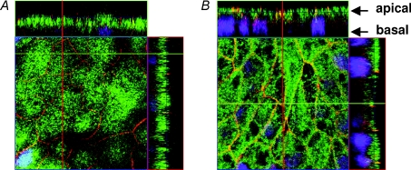
Figure 7. Localization of bestrophin in mock-transfected and claudin-16-transfected cells.
A, confocal laser-scanning micrographs of mock-transfected, and B, wt claudin-16 (short version)-transfected MDCK-C7 cells. Red, occludin; green, bestrophin; blue, nuclei (DAPI). The yellow colour along the cell–cell contacts in B indicates co-localization of occludin and bestrophin. This was not found in control cells. Z scans indicate that bestrophin accumulated not only at tight junctions but generally in the apical membrane.

