
Figure 2. Polyclonality of BCL6 transformed cell lines.
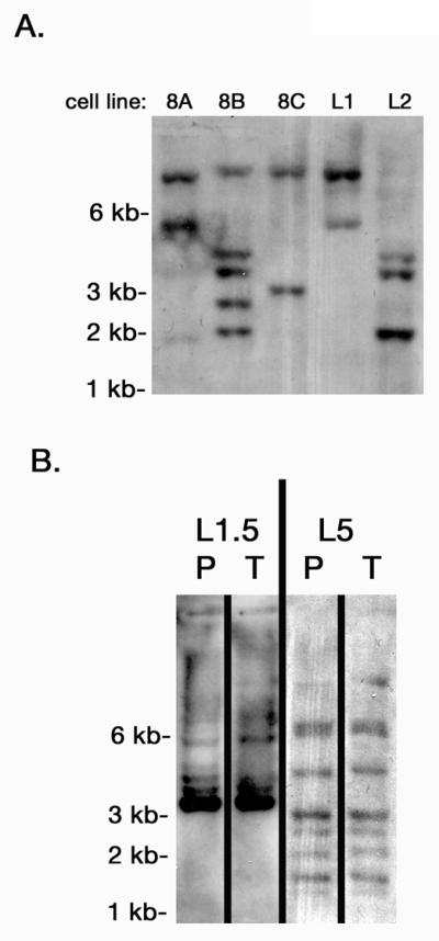
Southern blots with EcoRI-digested genomic DNA from the indicated BL B cell lines were probed with a 32P-labeled DNA fragment derived from the GFP gene of the pMIEG3-BCL6 vector. The different sized bands show different retroviral insertions representing unique B cell clones. A. Five different BL B cell lines. B. Two different BL B cell lines showing similar clones in the parental cell line (P) as in the cells isolated from a tumor (T) derived from the parental cell line.