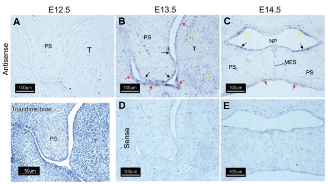
Fig. 1. Expression of Wnt-10a in the murine secondary palate.
Cryostat sections of fixed, frozen heads from mice on days E12.5–E14.5 were processed for expression of Wnt-10 transcripts by in situ hybridization with either an antisense Wnt-10a-specific riboprobe (A–C) or the sense (negative) control (D,E). An E12.5 coronal section stained with 0.05% (w/v) Toluidine Blue O is included for orientation. No expression of Wnt 10a mRNA was detected on E12.5 (A). On E13.5, expression of Wnt 10a was noted in both the future oral and nasal palatal epithelium and in the medial edge epithelium (B) (black arrows). Expression was also detected in the epithelium of the oral cavity and tongue (B, red arrows). Diffuse expression was observed in tongue mesenchyme on E13.5 (B, yellow arrows). Following shelf elevation and initiation of fusion on E14.5 (C,E), Wnt-10a expression was predominant in palatal nasal epithelium (C, black arrows) in the epithelium lining the nasopharynx (C, yellow arrows) and in the oral epithelium of the palate (C, red arrows). A positive signal was also observed in the medial edge epithelial seam (C, MES,). Abbreviations for all figures are: MES, medial edge seam; NP, nasopharynx; PS, secondary palatal shelf; T, tongue. All panels, 200X magnification.

