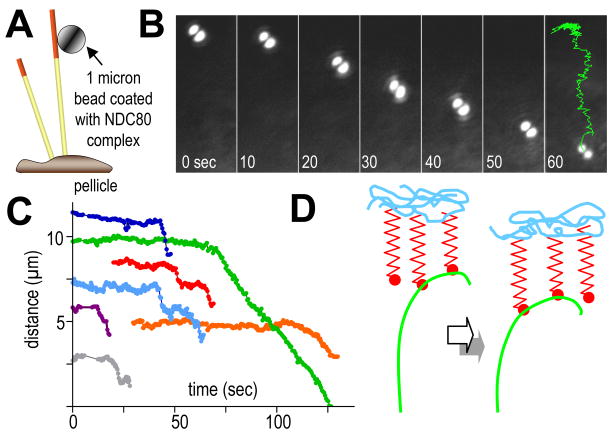
Figure 6. Beads coated with Ndc80 complex moving with depolymerizing MTs in vitro.
A. Experimental design (not to scale). Bovine brain MTs were capped with GMPCPP-assembled, rhodamine-labeled tubulin, so they would break up upon illumination with green light, permitting MT depolymerization.
B. Bead position at times shown (sec); last time shows trajectory of bead center for the entire motion.
C. Distances from bead to pellicle as a function of time for 7 separate experiments. Movement began shortly after the MT cap was removed.
D. Two consecutive times during the life of a PF from a depolymerizing MT that is stably attached to a “load”. The propagation of depolymerization, reflected in a progression of PF position, allows recycling KFs to transmit force to the load without a noticeable change in PF curvature where the KFs attach.

