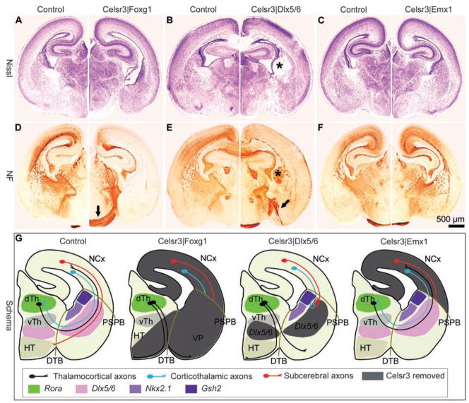
Fig. 2.
Region-specific Celsr3 inactivation affects development of the IC in different manners. (A to F) Montages of P0 sections stained with Cresyl violet [(A) to (C)] or neurofilament antibody (NF) [(D) to (F)]. The IC is fully defective in Celsr3|Foxg1 mice in which some thalamic axons cross to the contralateral diencephalon [arrow in (D)]. In Celsr3| Dlx5/6 mice, the IC does not form, but cortical axons stall and form a mass at the level of the striatum [asterisks in (B) and (E)], whereas thalamic fibers are mis-routed to the amygdala [arrow in (E)]. In Celsr3| Emx1 mice, the IC is present and thalamocortical connections are similar to that in normal mice [(C) and (F)]. (G) Schematic summary of the IC phenotypes in the various mice used, in relation to areas of Celsr3 inactivation (gray) and expression of markers (Dlx5/6, Gsh2, Nkx2.1, and Rora). dTh and vTh, dorsal and ventral thalamus; HT, hypothalamus; VP, ventral pallidum; NCx, neocortex; PSPB, pallial subpallial boundary; and DTB, diencephalon telencephalon border.

