
Fig. 4.
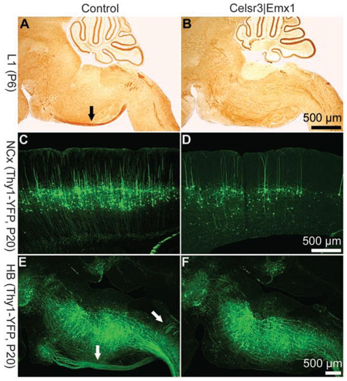
The corticospinal tract is defective in Celsr3|Emx1 mice. Comparison of control [(A), (C), and (E)] and Celsr3| Emx1 [(B), (D), and (F)] mice. In sagittal sections at P6 stained with an antibody against the L1 molecule (A) and (B), corticospinal axons are labeled in control [arrow in (A)] but not in the mutant ventral hindbrain. Crosses were carried out with Thy1-YFP mice, a transgene that labels neurons in cortical layer 5 and corticospinal axons (C) to (F). At P20, layer 5 is well populated in control mice (C), and the corticospinal tract is clearly defined [arrows in (E)], whereas cortical layer 5 is very diminutive (D) and no corticospinal axons are detected in the hindbrain (F) of Celsr3|Emx1 mice.