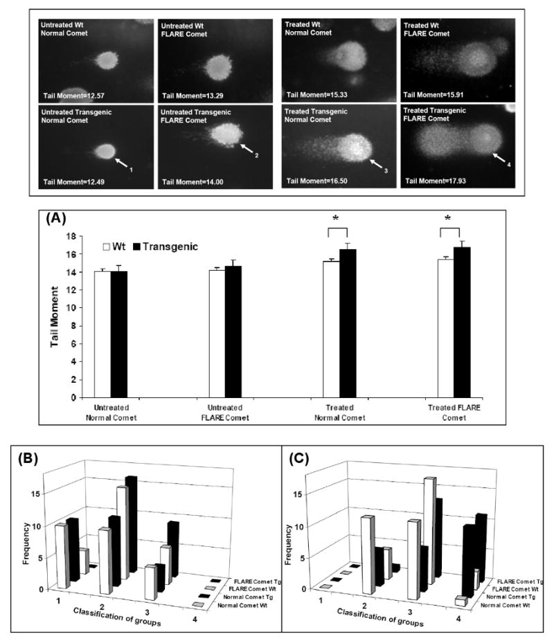
Figure 2. Increased incidence of oxidative DNA damage in transgenic cells after exposure to H2O2.

MEF wild-type cells or RPS3 transgenic cells were treated with 1 mM H2O2 for 48 h, and evaluated for the extent of DNA damage by normal comet assay and FLARE comet assay. (A) For each sample, tail moments expressed as adjusted tail moment (normalizing and variance-stabilizing logarithmic transformation applied to the calculated tail moments) of 25 cells from randomly selected fields were calculated and statistically significant differences determined by ANOVA analysis are represented with P-values. *, P=<0.01. DNA damage to individual cells was also determined by a visual scoring method [11] and plotted as cell frequency graphs where DNA damage to cells was divided into the following categories: type1- intact nucleus, smooth outer edges; type2- intact nucleus, small amount of tailing; type3- intact nucleus, large amount of tailing; type4- shrinking nucleus, large amount of tailing. (B) Unexposed wild-type and transgenic MEF cells show more number of cells with minimal damage (type1 and 2) for both normal comet and Fpg treated FLARE comet analysis. However in H2O2 exposed MEF cells we see a significant number of damaged cells (type 3 and 4) in transgenic MEF cells for both normal and FLARE comet compared to wild-type MEF cells (C).
