
Figure 4. In situ apoptosis detection in fixed tissue.
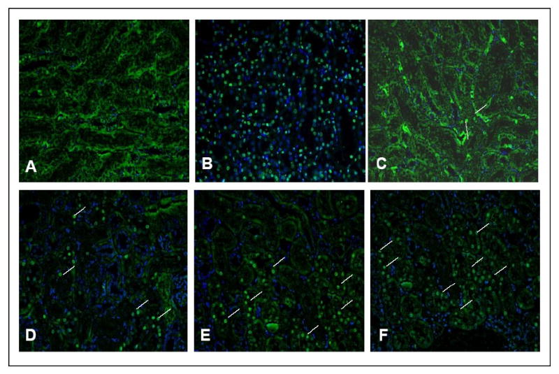
Kidney and liver tissue sections from age-matched 13-month-old wild-type and Tg (RPS3) male mice were labeled in situ to detect apoptotic cells in these tissues. DAPI was used to stain the nuclei. Merged images of FITC and DAPI channels that represent wild-type (panel C-D; different fields) and Tg (panel E-F; different fields) kidney respectively are shown. Apoptotic cells are indicated by white lines. Panel (A) is negative control where the TdT labeling enzyme was omitted in the staining process and panel (B) shows positive control treated with the TACS-nuclease to generate DNA strand breaks.